-
-
sample data

|
-
-
-
sample data

|
-
sample publication

|
|
BrainMaps Features the Highest Resolution Whole Brain Atlases Ever
Constructed
The resolution of BrainMaps data is 0.46 microns per pixel, with typical macaque brain image slices being over 100,000 pixels in width. This far exceeds the resolution found in print atlases, and is the highest resolution whole brain data to be found anywhere, either in print or online.
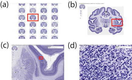
An example of navigation through virtual slides at brainmaps.org
using the Chlorocebus
aethiops (African Green Monkey) Nissl dataset. All images are
actual screenshots from a web browser and are what a visitor to
brainmaps.org would see. (A) An array of virtual slides for the
Chlorocebus aethiops dataset, shown as clickable thumbnails that, when
clicked on, launch a new browser window allowing navigation through the
high-resolution image (B). The image in (B) is 95,040 x 74,711 pixels
and 20 gigabytes in size. The thumbnail in the upper left is for
navigation purposes. Shown also are overlying labels of brain areas that
may be toggled on and off. (C) Zooming in on the slide in (B). The red
box in (B) corresponds to (C). (D) Zooming in to full resolution in (C),
showing details of individual neurons in the insula. The red box in (C)
corresponds to (D)
.
|
|